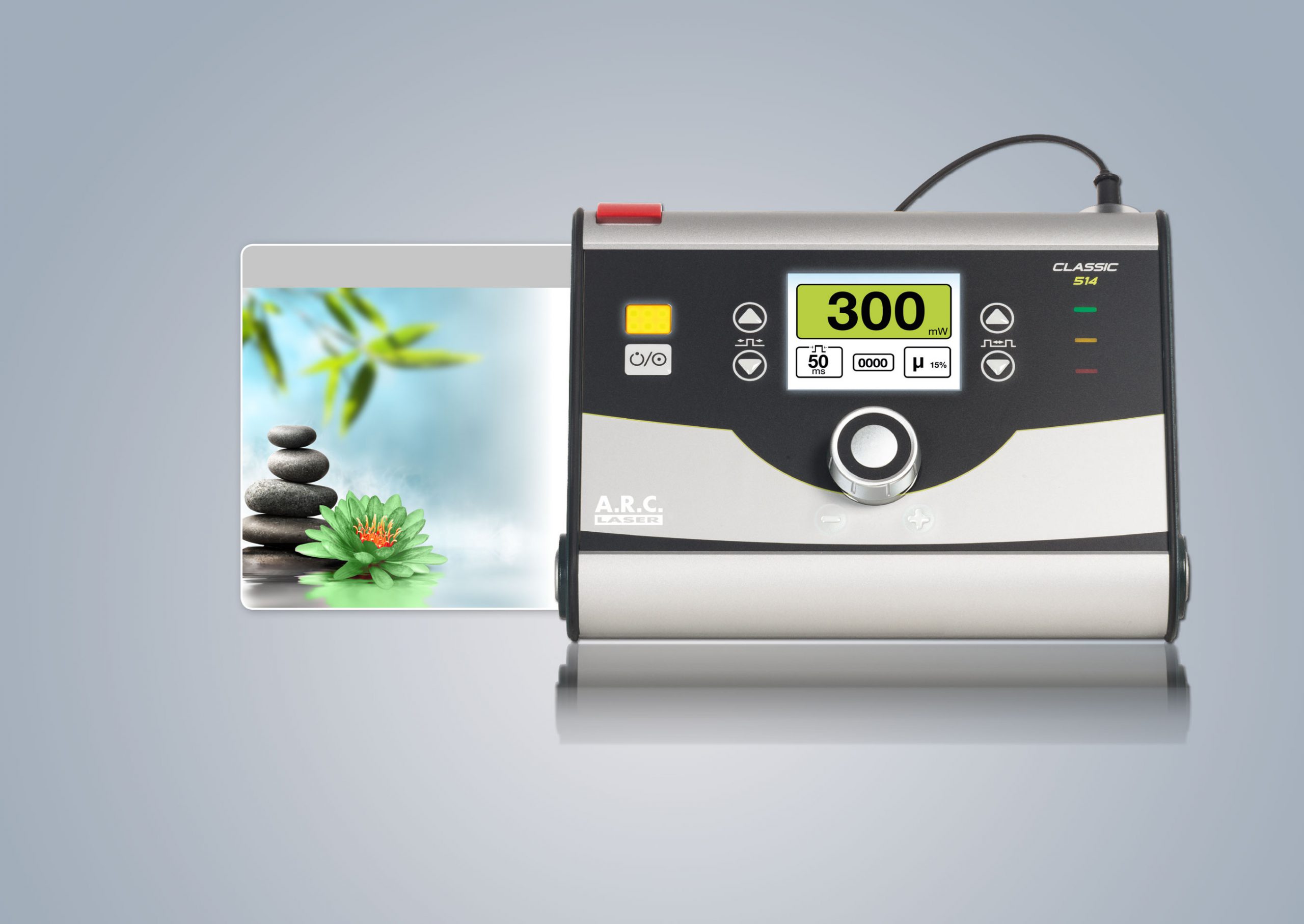
A.R.C LASER – CLASSIC 514 nm LASER VERDE ALLO STATO SOLIDO PORTATILE
13 Aprile 2020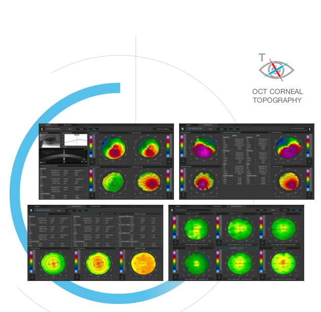
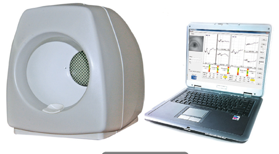
Moduli Aggiuntivi per OCT REVO: Topografo/Biometro Ottico/Angio OCT
13 Aprile 2020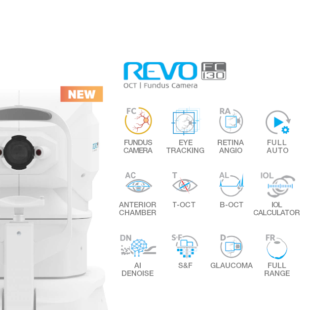
OPTOPOL – Copernicus REVO NX 130.000 Scan/Sec, REVO NX FC 130.000 Scan/Sec
l’OCT più veloce al mondo
Optopol engineering team, designers of the first commercially available Spectral Domain OCT in the World, is proud to present the World’s fastest OCT.
Optopol engineering team, the designers of the firstcommercially available Spectral Domain OCT in the world, are proud to present the latest innovation, the world`s first B-OCT and T-OCT for standard posterior OCT. Our supreme experience in Spectral Domain OCT allows us to provide the market with a state of the art instrument which comes with new advanced technologies and remarkable simplicity of operation.
FUNDUS CAMERA (ONLY IN FC MODEL)
A 12.3 MP Fundus Camera is integrated into our All in One OCT device capable of capturing detailed color images of ultra-high quality.
The REVO FC130 is fully automated, safe and easy to use.
- Color fundus image capture is possible with a pupil as small as 3.3mm.
- Easy to use fundus image processing tools deliver a stunning retinal image.
- Available modes deliver detailed photos of one or both eyes, as well as a chronological comparison of the fundus photo.
- Link a singe fundus photo to several OCT scans.
- IR fundus preview and photo capture settings are adjusted automatically based on the IR fundus preview.
- To meet the requirements of screening programs and allow the user to take exams for both eyes in non mydriatic mode, the device now has three auto flash levels.
| Retina 3D Both Eyes Retina View | Retina 3D Both Eyes Fundus View |
 |
 |
| Retina B-scan Single View | |
 |
The Revo FC130 is an All in One device you can use in a number of ways:
- as a Full Color Fundus Camera.
- as a combo providing simultaneous OCT and fundus images.
- for high quality OCT imaging including OCT-A.
- as a Biometry device.
The device offers all proven advantages of REVO systems with a cutting-edge color fundus imaging for a new level of diagnostic certainty. High quality OCT scanning and a comprehensive analysis of the retinal layers combined with a fundus imaging make the examination versatile as never before.
What makes the REVO FC130 truly unique is its integrated non-mydriatic 12.3 Mpix Fundus Camera capable of capturing ultra-high quality and detailed color images. The REVO FC130 Fundus Camera is fully automated, safe and easy to use.
- The advanced optical system ensures high quality imaging at a viewing angle as wide as 45°.
- New linking function makes it possible to link a single fundus photo to several OCT exams to reduce the number of shots per eye.
- Easy to use image processing tools such as RGB channel, brightness, contrast, gamma and sharpness adjusters used with filters deliver a stunning retinal image.
- Available view modes present detailed photos of a single or both eyes as well as a time comparison of fundus photos.
| Full screen Fundus Photo view | Both eyes Fundus Photo view |
 |
 |
OPTICAL COHERENCE TOMOGRAPHY
REVO FC130 with 130 000 A-scan/sec scanning speed utilizes state-of-the-art technologies and offers remarkable simplicity of operation. It meets all requirements for a modern OCT.
ACCUTRACKTM
Real time hardware eye tracking
The REVO FC130 now comes with a real-time hardware eye tracking function which compensates blinks, loss of fixation and involuntary eye movements during OCT scanning.
| ACCUTRACKTM |
The iTracking function is still available and proves useful while examining patients who find it difficult to maintain fixation.
AI DENOISETM
Improved tomogram quality powered by artificial intelligence. Advanced AI algorithms enhance the quality of a single tomogram to the level of an averaged tomogram obtained through multiple scans. The AI DeNoise algorithm filters out noise from the tomogram for the highest and smoothest image quality. The function is available on all tomograms and in every tab featuring them, including the 3D tab. On averaged tomograms the function is on by default. The moment a tomogram is loaded for review the software starts denoising it. After a short moment the original “undenoised” tomogram is replaced with a noise-free image.
AiDenoise Tomogram |
AiDenoise Tomogram |
 |
 |
FULL RANGE
With scans presenting New Extended DepthTM software, based on our Full Range technology, provides scans of increased depth for reliable and convenient observation of challenging cases. With scans presenting extended depth, this new imaging mode is perfect for diagnosing even highly myopic patients. 
RETINA
A single 3D Retina examination is sufficient to perform both Retina and Glaucoma analysis based on retinal scans. During the analysis the software automatically recognizes 8 retina layers to ensure a more precise diagnosis and mapping of any changes in the patient’s retina condition.
| 3D | |
 |
|
| 3D Retina + Fundus Photo Single View | Both VIew |
 |
 |
FOLLOW UP
High density of standard 3D scan allows the operator to precisely track the disease progression. The operator can analyze changes in morphology, quantified progression maps and evaluate the progression trends.
Precise registration
The software can track 3D scans and register them to the OCT baseline exam by recognizing patterns in the shape of blood vessels. Active tracking and post-processing point-to-point registration allows the user to precisely see and track the changes in retina morphology in Comparison and Progression analysis.
| Progression Morphology | Progression Quantification |
 |
 |
| Comparsion |
 |
Auto Flash
IR fundus preview and the photo capture settings are adjusted automatically based on the IR fundus preview. This ensures correct automatic flash setting for perfect images regardless of the pupil size and eye pigmentation.
No need to re-take images because of wrong flash settings.
Fundus photograph intensity mode
By selecting the flash mode the operator can determine the quality level suitable for obtaining a detailed image or a screening photograph. This means that in many cases the exam time and miosos can be reduced to increase patient comfort.
Fundus photograph screening
To meet the requirements of screening programs and allow the user to take exams of both eyes in non mydriatic mode, the device now has three auto flash levels. Using the Flash Low setting, it is possible to take pictures of both eyes in less than 25 seconds.
Anterior segment photo
This new mode allows the user to take color photos of the anterior segment, presenting the cornea, eyelid, pupil and sclera.
| Anterior Photo Eye View | Anterior Photo Eye View |
 |
 |
OCT ANGIOGRAPHY
This module allows visualization of the retinal microvasculature. Angiography SOCT is a non-invasive, dye-free technique providing 3D image of retinal blood circulation.
| Standard Single View | Detailed Single View |
 |
 |
| Comparison View | Progression |
 |
 |
ANGIO ANALYISIS METHODS
QUANTIFICATION
The quantification tool provides quantification of the vasculature in the whole analyzed area together with values in specific zones and sectors. Thanks to the heat map of the analyzed vasculature the evaluation of vascular structure conditions is much faster. The choice of the quantification method increases the sensitivity of analyses for specific deseases.
Available quantification methods:
- Vessel Area Density – it is defined as the total area of perfused vasculature per unit area in a region of measurement.
- Skeleton Area Density – it is defined as the total area of skeletonized vasculature per unit area in a region of measurement.
Quantification is available for a specific layer in Angio OCT exam:
- Retina: Superficial Plexus and Deep Plexus.
- Disc: RPC – Radial Peripapillary Capillary.
 |
 |
| Skeleton Density Map | Vessel Density Map |
ANGIO-ANALYTICAL TOOLS
FAZ – Foveal Avascular Zone measurements enable the quantification and monitoring of changes in Superficial and Deep vascular layers. The FAZ tool is also available for narrow and wide scans.
 |
 |
| FAZ Area [mm2] Perimeter [mm] Circularity |
VFA – Vascular Flow Area allows the user to examine the pathologically affected area and to precisely measure the area covered by vascularization.
The simple and easy area measurement can be performed on a predefined or user-selected vascular layer.
 |
 |
| Area [mm2] Flow Area [mm2] |
NFA – Non Flow Area measurement tool makes it possible to quantify the Non Flow Area on the OCT Angio examination. It provides the sum of all marked areas.
 |
 |
| Non Flow Area [mm2] |
ANGIO MOSAIC
The Angiography mosaic delivers high-detailed images over large field of the retina. Advanced tab provides: view of any vascular layers, enface view of vascular layers, depth coded and thickness map.
| Mosaic modes: 7x7mm | PDR Angio Mosaic mode: 10x10mm |
 |
 |
GLAUCOMA
Comprehensive glaucoma analytical tools for quantification of the Nerve Fiber Layer, Ganglion layer and Optic Head with DDLS enable the user to perform precise diagnosis and monitoring of glaucoma over time.
With the golden standard 14 optic nerve parameters and a new Rim to Disc and Rim Absence the description of ONH condition is quick and precise.
Advanced view which provides combined information from Retina and Disc scan to integrate details of the Ganglion cells, RNFL, ONH in a wide field perspective for comprehensive analysis.
Advance Retina & ONH |
ONH Single |
 |
 |
Asymmetry analysis of Ganglion layers between hemispheres and between eyes helps to detect and identify glaucoma in early stages and in non-typical patients.
Ganglion Both |
Ganglion Progression |
 |
 |
The REVO DDLS (Disc Damage Likelihood Scale) uses 3 separate classifications for small, average and large discs. It supports the practicioner in a quick and precise evaluation of the patient’s glaucomatous disc damage.
ONH Both |
ONH Progression |
 |
 |
COMPLETE YOUR GLAUCOMA REPORT
To eliminate the common problem of understanding the patient’s IOP, the pachymetry module provides IOP Correction value. With
the implemented Adjusted IOP formula, you can quickly and precisely understand the measured IOP value. The Pachymetry and
Anterior Chamber Angle Verification require no additional attachments. The predefined Glaucoma protocol, which consists of Retina,
Disc and Anterior scans, can be done automatically to reduce patient chair time.
Closed angle |
Full Range Anterior Chamber |
 |
 |
| *image courtedy of Bartosz L. Sikorski PhD | *image courtedy of Bartosz L. Sikorski PhD |
| Anterior Radial Single View Cornea | |
 |
|
| *image courtedy of Bartosz L. Sikorski PhD | |
COMPREHENSIVE GLAUCOMA SOLUTION
STRUCTURE & FUNCTION – Combined OCT and VF results analysis
COMPRHENSIVE STRUCTURE AND FUNCTION REPORT INCLUDES THE FOLLOWING
- VF sensitivity results
- 24-2/30-2 or 10-2
- Total and Pattern Deviation probability graphs for VF results
- Reliability and Global indices for VF results
- Combined map of Structure & Function
- Ganglion cells analysis (GCL+IPL or NFL+GCL+IPL)
- ONH and NFL analysis including charts and comparison tables
- NFL Asymmetry Plot
- Nasal and Temporal sectors have been spilt to present structural changes better
- Compare exact numerical sensitivity values
| Structure & Function |
 |
SINGLE PAGE REPORT
S+F provides a quick and comprehensive single page report for glaucoma management.
ANTERIOR CHAMBER
Built-in anterior lens allows the user to perform the imaging of the anterior segment without installing additional lens or forehead adapter. Now you can display the whole anterior segment or focus on a small area to bring out the details of the image.
Anterior Chamber exam with a fast view of the whole Anterior Chamber make the evaluation of gonioscopy situation and the verification of cataract lens easier and faster.
| Full Range Anterior Chamber | Full Range Anterior Chamber with Lens |
 |
 |
| Full Range Lens | |
 |
ANTERIOR SEGMENT
See incredible details of a selected area of the anterior segment.
| Cornea Single | Cornea Both |
 |
 |
| Cornea Comparison | Cornea Progression |
 |
 |
BIOMETRY OCT
B-OCT® is an innovative method of using the posterior OCT device to measure ocular structure along eye axis.
OCT Biometry provides a complete set of Biometry parameters: Axial Length AL, Central Cornea Thickness CCT, Anterior Chamber Depth ACD, Lens Thickness LT, Pupil size P and White to White WTW.
The B-OCT® module is available in two options:
- Standard Biometry module:
- High myopia management
- Axial distance measurement
- IOL Calculator
- IOL lens editor
- IOLcon.org support
The standard version of the module broadens the functionality of the device with the addition of the IOL Tab which allows the user to calculate the lens power of a selected IOL model (on the basis of the patient’s morphological data).
- Basic Biometry module:
- High myopia management
- Axial distances measurement
- No IOL Calculator
- Optometrist dedicated module
The basic B-OCT® module provides an exceptional comfort of daily use.
Result review |
Analysis window |
IOL CALCULATOR
IOL formulas allow the user to calculate IOL implant parameters. Our systems now support the latest IOL data base standard IOLCon.org so that you can always keep your library up-to-date.
CORNEAL TOPOGRAPHY
The corneal topography module shows the changes in the cornea on the diff erence map view. Customize favored view by selecting from a variety of available maps and display options. Fully Automatic Capture with examination time of up to 0.2 sec makes testing quick and easy.
- Full featured Corneal mapping of Anterior, Posterior and Real Precise Astigmatism Display Option (SimK: Anterior, Posterior, Real, Meridian and Semi-Meridian ø 3, 5, 7 mm zones)
Easily detect and classify keratoconus with the Keratoconus classifier. Classification based on KPI, SAI, DSI, OSI and CSI. In the early stages of keratoconus the results can be complemented by Epithelium and Pachymetry maps.
Normal |
Astigmatysm |
Keratoconus |
 |
 |
 |
COMPARE THE TOPOGRAPHY EXAMS
Comprehensive software features a range of selectable views: Single, Both. The user can observe details on the standard Singe view and easily see corneal asymmetry on the Both view.
The follow-up feature in the T-OCT™ module gives the possibility to fully compare the changes in the corneal topography over time for:
- LASIK undergone patients
- Keratoconus patients
- The contact lens wearers
| Single | Both |
 |
 |
| Comparison | Progression |
 |
 |
NETWORKING
A proficient networking solution increases productivity and enhances patient experience. It allows you to view and manage multiple examinations from review stations in your practice. It also effortlessly facilitates patient education by allowing you to interactively show examination results to patients. Every practice will have different requirements which we can cater for by tailoring a bespoke service. There is no additional charge for the server module.
DICOM
Receiving Worklist (MWL), Store, exchange and transmit results via through DICOM storage gateway to the hospital network.
| Marca |
|---|