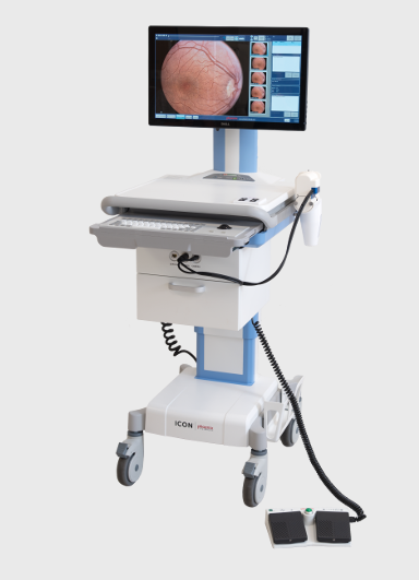
PHOENIX CLINICAL – ICON Strumento di immagini a grande campo per pazienti pediatrici
2 Agosto 2018
OPHTEC – PRECIZON PRESBYOPIC
2 Novembre 2018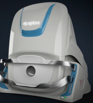
OPTOS – MONACO
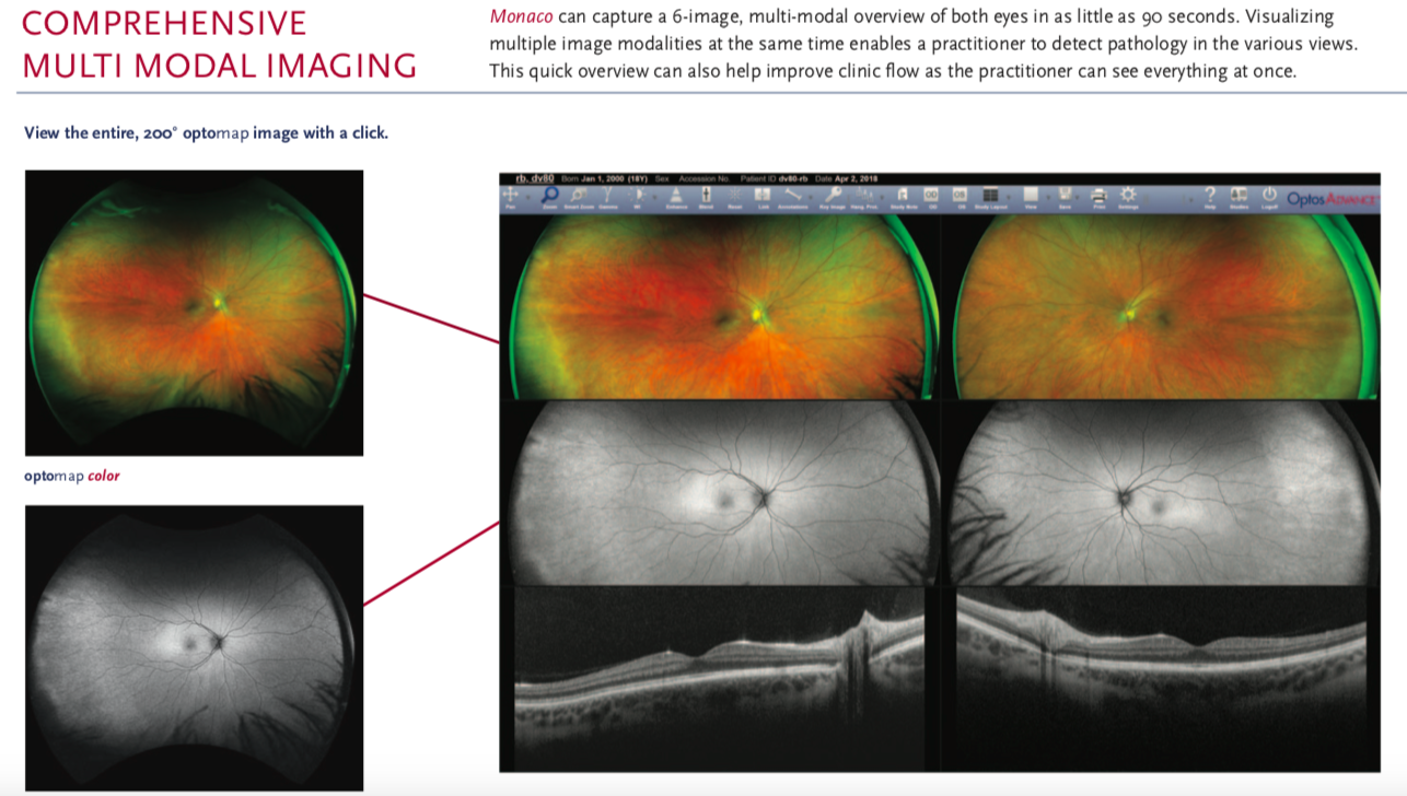
Monaco is an exciting new way to enhance your clinical exam. The only ultra-widefield retinal imaging device with integrated OCT, Monaco produces a 200° single-capture optomap image in less than ½ second and also provides cross-sectional 40° OCT views of retinal structures. Monaco enables a rapid multi-modality capture featuring color, autofluorescence and OCT scans, for both eyes, in as little as two minutes.
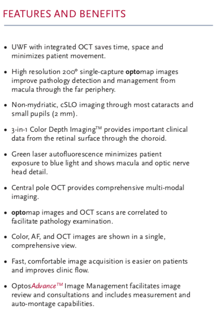
Monaco offers the following benefits:
• UWF with integrated OCT saves time, space and minimizes patient movement
• Central pole OCT provides comprehensive multi-modal imaging
• optomap images and OCT scans are correlated to facilitate pathology examination
• Color, AF, and OCT images are shown in a single, comprehensive view
VIDEO:
Technical Specifications
| Image Modalities | optomap color and optomap plus (red and green laser): Color composite view Green laser view Red laser view optomap af (green laser): autofluorescence Optical Coherence Tomography (OCT) |
|---|---|
| Resolution | optomap color: 20 μm optomap plus, af : 14 μm |
| Wavelengths | Red laser: 635 nm green laser: 532 nm (for af ) |
| Exposure Time | Less than 0.4 seconds |
| Tomographic Imaging | Signal Type: Optical scattering from tissue Signal Source: Super luminescent Diode (SlD) 830 nm Optical Power: laser safety Class-1 following IEC/en60825-1:2014 Typical Axial Resolution: <10 micron (in tissue) Digital on-screen <6 micron Transverse Resolution: 20 micron (in tissue) Scanners: Galavanometric with x, y mirrors Scan Depth: Up to 2.5mm |
| OCT Scan Characteristics | Spectral Domain OCT A-Scan rate up to 70k cycles/s Active eye tracking Automatic scan positioning |
| OCT Scan Types | Line Scan Raster Scan Retina Topography Scan Optic nerve Head (OnH) Topography Scan Retinal nerve Fiber layer (RnFl) Scan |
| Footprint | Width: 550 mm/22 inches Depth: 500 mm/19.5 inches Height: 650 mm/25.5 inches |
| Marca |
|---|